קובץ:Fundus of patient with retinitis pigmentosa, mid stage.jpg
גודל התצוגה המקדימה הזאת: 699 × 599 פיקסלים. רזולוציות אחרות: 280 × 240 פיקסלים | 560 × 480 פיקסלים | 871 × 747 פיקסלים.
לקובץ המקורי (871 × 747 פיקסלים, גודל הקובץ: 107 ק"ב, סוג MIME: image/jpeg)
| זהו קובץ שמקורו במיזם ויקישיתוף. תיאורו בדף תיאור הקובץ המקורי (בעברית) מוצג למטה. |
| תיאור |
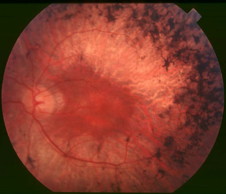
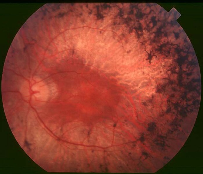
English: Figure 2. Fundus of patient with retinitis pigmentosa, mid stage (Bone spicule-shaped pigment deposits are present in the mid periphery along with retinal atrophy, while the macula is preserved although with a peripheral ring of depigmentation. Retinal vessels are attenuated.) Hamel Orphanet Journal of Rare Diseases 2006 1:40 doi:10.1186/1750-1172-1-40 |
| תאריך יצירה | |
| מקור | Retinitis pigmentosa by Christian Hamel |
| יוצר | Christian Hamel |
| אישורים והיתרים (שימוש חוזר בקובץ זה) |
© 2006 Hamel; licensee BioMed Central Ltd. This is an Open Access article distributed under the terms of the Creative Commons Attribution License (https://creativecommons.org/licenses/by/2.0), which permits unrestricted use, distribution, and reproduction in any medium, provided the original work is properly cited. |
הקובץ הזה מתפרסם לפי תנאי רישיון קריאייטיב קומונז ייחוס 2.0 כללי.
- הנכם רשאים:
- לשתף – להעתיק, להפיץ ולהעביר את העבודה
- לערבב בין עבודות – להתאים את העבודה
- תחת התנאים הבאים:
- ייחוס – יש לתת ייחוס הולם, לתת קישור לרישיון, ולציין אם נעשו שינויים. אפשר לעשות את זה בכל צורה סבירה, אבל לא בשום צורה שמשתמע ממנה שמעניק הרישיון תומך בך או בשימוש שלך.
היסטוריית הקובץ
ניתן ללחוץ על תאריך/שעה כדי לראות את הקובץ כפי שנראה באותו זמן.
| תאריך/שעה | תמונה ממוזערת | ממדים | משתמש | הערה | |
|---|---|---|---|---|---|
| נוכחית | 13:17, 2 בדצמבר 2017 | | 747 × 871 (107 ק"ב) | Doc James | Cropped 27 % horizontally and 7 % vertically using CropTool with precise mode. |
| 16:52, 22 בספטמבר 2009 |  | 799 × 1,200 (126 ק"ב) | CopperKettle | {{Information |Description={{en|1=Figure 2. Fundus of patient with retinitis pigmentosa, mid stage (Bone spicule-shaped pigment deposits are present in the mid periphery along with retinal atrophy, while the macula is preserved although with a peripheral |
שימוש בקובץ
הדף הבא משתמש בקובץ הזה:
שימוש גלובלי בקובץ
אתרי הוויקי השונים הבאים משתמשים בקובץ זה:
- שימוש באתר ar.wikipedia.org
- שימוש באתר bs.wikipedia.org
- שימוש באתר ca.wikipedia.org
- שימוש באתר da.wikipedia.org
- שימוש באתר en.wikipedia.org
- שימוש באתר en.wikiversity.org
- שימוש באתר es.wikipedia.org
- שימוש באתר eu.wikipedia.org
- שימוש באתר fa.wikipedia.org
- שימוש באתר fi.wikipedia.org
- שימוש באתר fr.wikipedia.org
- שימוש באתר hy.wikipedia.org
- שימוש באתר it.wikipedia.org
- שימוש באתר ko.wikipedia.org
- שימוש באתר la.wikipedia.org
- שימוש באתר or.wikipedia.org
- שימוש באתר outreach.wikimedia.org
- שימוש באתר pl.wikipedia.org
- שימוש באתר pt.wikipedia.org
- שימוש באתר ru.wikipedia.org
- שימוש באתר sl.wikipedia.org
- שימוש באתר sr.wikipedia.org
- שימוש באתר sv.wikipedia.org
- שימוש באתר th.wikipedia.org
- שימוש באתר tr.wikipedia.org
- שימוש באתר tt.wikipedia.org
- שימוש באתר uk.wikipedia.org
- שימוש באתר vi.wikipedia.org